

Nelson Jen An Chao, MD
- Professor of Medicine
- Donald D. and Elizabeth G. Cooke Cancer Distinguished Research Professor
- Professor in Immunology
- Research Professor of Global Health
- Professor in Pathology
- Member of the Duke Cancer Institute
- Chief, Division of Cell Therapy in the Department of Medicine
- Affiliate of the Regeneration Next Initiative

https://medicine.duke.edu/faculty/nelson-jen-chao-md
Similac Neo-Care blood pressure cuff cvs purchase plendil without a prescription, designed for containing docosahexaenoic acid and arachidonic acid have preterm infants weighing more than 1800 g at birth blood pressure chart high systolic low diastolic plendil 5 mg line, provides been recently marketed to promote eye and brain develop 22 cal/oz in standard dilution pulse pressure 95 order plendil discount. So far no randomized trials have shown any benefit although no harm has been established blood pressure table order cheap plendil line. There are also designed to supplement calories hypertension obesity buy discount plendil online, protein heart attack pain in left arm cheap 2.5 mg plendil with visa, phosphorus, cal multiple lactose-free preparations. Increased osmolality may enhance gastrointestinal irritability and affect tolerance. Because the proteins are plant Similac Natural Care is a liquid milk fortifier that is typi based, vitamin and mineral composition is increased to com cally mixed in a 1:1 ratio with breast milk. Other alternatives pensate for plant-based mineral antagonists while supple may include feedings with breast milk and fortifier. The menting protein composition with the addition of osmolality of Similac Natural Care is lower than that of methionine. This liquid fortifier may be prefer owing to a carbohydrate composition that includes sucrose able, particularly for infants whose mothers have low milk and corn syrup. Soy-based formulas should Other specially formulated formulas are available includ not be used for preterm infants because they cause less ing antireflux and hypoallergenic formulas. Reflux usually weight gain and increase the risk of osteopenia of prematu does not require treatment unless there is poor weight gain. ProSobee, Isomil, and I-Soyalac are common soy-based Antireflux formulas decrease emesis and regurgitation, but preparations. This formula, which contains American Dietetic Association: Nutrition Management of the casein-based protein and glucose, is not recommended for Infant. Pediatrics weight, and with three times the vitamin and mineral con 1994;94:547-549. Premature infant preparations are Black R, Jarman L, Simpson J: Silicone breast implants and breast approximately 60% casein and 40% whey, with 1:1 concen feeding. J delivery on duration of breastfeeding: a prospective cohort Obstet Gynecol Neonatal Nurs 2003;32:40. Breastfeed Thygarajan A, Burks A: American Academy of Pediatrics recom Rev 2002;10:19. Resources for breastfeeding products and information: Pediatrics 2002;110(5):1030. Baraff and colleagues published a set of useful prac common types of problems encountered by physicians who tice guidelines that are summarized in Table 5-1. The frequency and nature of serious bacterial illness is different in the three different age groups. However, existing screening protocols lack the sensitivity and negative General Considerations predictive value to identify infants at low risk for these infec Fever is the primary sign that indicates an infectious process tions. For this reason, it is generally accepted that all febrile in children of all ages. Other than fever, however, many chil infants younger than 1 month of age be admitted to the hos dren do not display signs or symptoms indicative of the pital, given a complete sepsis workup, and treated with par underlying disease. Twenty percent of febrile children, after enteral antibiotics pending the results of the workup. Of history and physical examination, have fever without a these infants, approximately 65% have a viral infection, 13% source of infection. Three percent have bacteremia, stool, as well as abscess or cellulitis and pneumonia with pos with group B Streptococcus, Enterobacter, Listeria, itive blood cultures. Children are generally divided into three Streptococcus pneumoniae, E coli, Enterococcus, and Klebsiella groups for evaluation purposes: young children aged 3 all being found. Fewer than 2% have meningitis, usually months to 3 years, young infants aged 2-3 months, and caused by Klebsiella, Listeria, and group B Streptococcus. There is no absolute demarca In evaluating infants older than 1 month of age, it is use tion between these ages. Rather, one age group fades into the ful to first identify which infants are at low risk for a serious next, and the physician is left to make a judgment about how bacterial illness. The criteria for low risk are being previously to treat each child in the border ages. Infant <<1 mo of age Child 3 mo to 3 y of age Admit for evaluation and treatment Toxic: admit Nontoxic: Infant 2-3 mo of age Temperature <<39. Source: Adapted from Baraff L: Management of fever without source in infants and children. The most common organ should be hospitalized and treated with parenteral antibiotics. The most common serious bacterial ill ratory infection, such as cough, tachypnea, rales, or rhonchi. The use of empiric antibiotics single intramuscular dose of ceftriaxone should be given. The child should be reevaluated in 18-24 hours and a second There is general consensus that bacteremia is a risk factor for dose of ceftriaxone given. If blood cultures are found to be development of infectious complications, such as meningitis. However, pneumococcal bacteremia responds well to oral If the urine culture is positive and the child has a persistent antibiotics, so these drugs can be used in children who fever, the child should be admitted for treatment. Table 5-1 presents guidelines that may be useful for inves tigating and treating febrile children. Clinical Findings Baraff L: Management of fever without source in infants and chil A. A careful, complete physical examination is necessary to exclude focal signs of infection. Bulging, immobile tympanic membrane that is dull gray, the abdomen should be examined for signs of peritonitis or yellow, or red in color. A musculoskeletal examination should be done Perforated tympanic membrane with purulent drainage looking for evidence of osteomyelitis or septic arthritis. Almost all children have at least one episode of otitis If the child has diarrhea, stool cultures should be evaluated. Treatment Pathogenesis All infants younger than 1 month of age should be hospital When cultures of middle ear fluid are done, S pneumoniae is ized. An appropriate antibiotic regimen includes ceftriaxone found in about 35%, H influenzae in about 25%, and (50 mg/kg/d) with or without gentamicin. Ten percent of effusions cillin has been used routinely to cover the possibility of show more than one of these bacteria, and about 25% are Listeria infection. Viruses are recovered in a large percentage of cases, infection with Listeria is decreasing, ampicillin may be added with or without bacteria, but whether their role is causative to this regimen if the physician chooses. The bacteria responsible for There are several identified risk factors for otitis media, not hematogenous spread are principally S pneumoniae and H all of which are easily modifiable for prevention of the dis influenza. Other risk factors Nonsuppurative complications are primarily those that include increased number of siblings in the house, exposure arise from middle ear effusion and inflammation and scar to tobacco smoke, pacifier use, formula feeding, and lower ring of the structures of the middle ear. Children with abnormalities of the does not influence the persistence of middle ear effusions palatal architecture, such as those with cleft palate or Down after otitis media, nor does it have any effect on long-term syndrome, are at greatly increased risk. In summary, it appears vaccines against H influenzae type b and S pneumoniae are that complications of otitis media may not be preventable by not expected to have much impact on the disease, as the antibiotic treatment. Symptoms and Signs symptoms within 24 hours without treatment, and between Despite the frequency with which physicians see children with 80% and 85% recover in 1-7 days without antibiotics. Narrow-spectrum antibiotics have the develop over only a few hours, or the onset may be more same success rate as broad-spectrum antibiotics, although gradual. Younger adverse effects, primarily gastrointestinal, are more common children do not localize pain as obviously as older children. All guidelines recommend oral amoxicillin as Fever is present only in about 25% and is more common in first-line therapy. The tympanic membrane bulges and may Pediatrics/American Academy of Family Physicians) guide be cloudy, yellow, or red in color. Erythema of the tympanic line recommends high-dose amoxicillin (80-90 mg/kg/d), as membrane may be caused by fever or by screaming, so this this dose has been found to be more effective against peni sign is of questionable reliability. The However, the studies supporting high-dose therapy are based infection is bilateral in half of affected children. The tympanic on bacteriologic cure; evidence that high-dose therapy is membrane ruptures in fewer than 5% of cases, but pus drain clinically superior is lacking. Many of the symptoms are mend azithromycin, trimethoprim-sulfamethoxazole, eryth identical, and findings in the tympanic membrane may be romycin, or cefaclor, except in cases of severe penicillin subtle and nondiagnostic. Studies also document that a 5-day course of antibiotics is as effective as the standard 10-day course. This may be considered in children the bacteria from the middle ear, primarily sepsis and menin over the age of 2 years if the presenting illness is not severe gitis. Adenoviruses can cause pharyngoconjunctival fever, with It is important to note that studies have not adequately exudative pharyngitis and conjunctivitis. Epstein-Barr virus addressed the issues of treatment of children younger than 2 causes infectious mononucleosis, which commonly produces years of age and treatment of frequently recurrent or com other signs, such as generalized lymphadenopathy and plicated otitis media. Physicians are left to their clinical judg splenomegaly, in addition to exudative pharyngitis. Herpesviruses and coxsackie viruses can cause ulcerative the best treatment for children with frequent recurrences stomatitis and pharyngitis. The that children will only benefit from daily antibiotic prophy literature contains numerous recommendations for diagno laxis if they have had more than three episodes in 6 to 18 sis and treatment, but there is no clear consensus as to the months. The magnitude of benefit is small, with a reduction most accurate or most cost-effective method for evaluation of about one episode per year. Group A -Hemolytic sistent otitis media with middle ear effusions has not been Streptococcal Infection found to improve developmental outcomes. Moderate to severe tender anterior cervical lym the half-life of the middle ear effusion is about 4 weeks, with 10% persistence at 4 months. American Academy of Family Physicians, American Academy of Pediatrics: Diagnosis and Management of Acute Otitis Media. Clinical Findings Takata G et al: Evidence assessment of management of acute otitis media: I. If all four of these are present, the likelihood of responsible for about 15% of cases of pharyngitis. Excluding scarlatiniform rash, the presence Antibiotic treatment has only a modest effect on the course of the remaining three gives a probability of greater than of the disease, but adequate treatment with antibiotics effec 65%. In the absence of moderate to severe tonsillar enlarge tively prevents the important complication of rheumatic fever. However, penicillin V is still considered the drug of choice for children who are not Rapid antigen detection tests are commonly used in practice. There is no agreement as to the best Although the sensitivities of these assays may be reported as alternative in penicillin-allergic children. Because of increas very high in laboratories, in practice they may have a false ing resistance to erythromycin in some areas, clindamycin is negative rate as high as 20%. It is well established that 10 days of throat culture is dependent on technique and may also have treatment is necessary to achieve the maximum possibility of a false-negative rate of 10%-20%. However, for reasons tor is the inability of either rapid antigen testing or culture to that are unclear, streptococci persist in the pharynx in about distinguish between a true streptococcal infection and a viral 10% of treated children, regardless of which antibiotic is used. Morbidity and mortality are primarily related to the previously mentioned complications. The nonsuppurative Attia M et al: Multivariate predictive models for group A beta complications are rheumatic fever and post-streptococcal hemolytic streptococcal pharyngitis in children. Treatment of the acute infection may shorten the course of the disease by a small amount, although untreated disease General Considerations will resolve within several days in most children. It is not clear whether immediate antibiotic treatment offers greater Peritonsillar abscess is the most common deep space head benefit than symptomatic treatment. It is generally believed and neck infection in children, accounting for almost half of that treatment reduces the rate of suppurative complications, these infections. The exact cause is unknown, but it is thought the initial streptococcal infection would not have been rec that the infection usually spreads from the tonsil itself into ognized in its earlier stages. For reasons that are unclear, numerous stud adults, but it affects older children and adolescents more ies show that cephalosporins have higher rates of clinical and than younger children. Symptoms and Signs Children generally recover uneventfully once appropriate treatment is begun, but they may be at increased risk for a Most children with peritonsillar abscess have had symptoms second infection. Many of these children have been treated with antibiotics for pharyngitis before developing the Schraff S et al: Peritonsillar abscess in children: a 10-year review of diagnosis and management. Computed tomography and ultrasound studies of the neck often show the abscess, but the diagnosis is generally made by General Considerations history and physical examination.
As for the arteries arteria hepatica propia order genuine plendil on line, the cially modified could reverse the arterial or venous shaping of the venous system follows chronologi character of the channels blood pressure chart 15 year old generic 10mg plendil mastercard, and even change the cally the development of the brain metabolism blood pressure simulator generic 10 mg plendil free shipping. As Streeter described it pulse pressure equivalent purchase plendil 2.5mg with mastercard, the vascular system in the cranial neural crest cells give the forebrain 3 weeks 5 and 6 is confined to the meninx primitiva blood pressure medication and zinc buy cheap plendil 2.5 mg on-line. The pericerebral meshwork divides into a deep A last point that should be mentioned regarding (futurepial) layeroverthebrain surfaceandasuper the brain arteries concerns the origin of their ficial (future dural) layer arrhythmia institute order plendil 5 mg visa. The deep layer is a simple smooth muscle cells and pericytes (cells that capillary layer. In the superficial layer channels mediate vasoconstriction and form part of the become continuous and connect with the paired blood-brain barrier). Like that of the meninx primi aorta and the cardinal veins, to form early arterial tiva, this origin is different in the cord, hindbrain, and venous trunks. Connections between the and midbrain vessels on the one hand, and in the superficial layer and the deep capillary layer forebrain vessels on the other hand. In the caudal become the arterial feeders and the venous drain parts of the brain and in the cord the arterial media ing channels (early form of bridging veins). By contrast, in the forebrain specialized and, to better support the brain tissue, they originate from the neural crest (or mesecto 1,6,7,20 33 develops the intraventricular choroid plexus. The forebrain itself originates from the Specific feeding and draining channels develop anterior neural plate, which forms an expansion accordingly. As mentioned earlier, venous drainage has been shown to run succes the skeleton of the face and anterior skull base sively through a ventral diencephalic vein toward (maxillary, nasal, orbital regions, paranasal the primitive transverse sinus and then, more sinuses) as well as the coverings of the brain importantly, through prominent bilateral dorsal (meninges, calvarium, scalp, dermis) cannot have 17 choroid veins. The main collector of the superior a somitic origin, and instead are produced by the choroid veins is a single, median dorsal vein called neural crest. As the anterior neural plate itself is the vena mediana prosencephali or median pros also devoid of neural crest, the midfacial skeleton encephalic vein (Fig. In a similar way, the same however that such a term was confusing, as both groups of neural crest cells form the muscular the tributaries and the course of this single median media and the pericytes of the forebrain arteries 34 meningeal vein are different from those of the later as well. The limit between the neural crest-asso 17 true, paired, choroidal internal cerebral veins. The ciated forebrain arteries and the classic endothe vein of Markowski is a single, median, dorsal pros lium-associated mid and hindbrain arteries is encephalic vein that is not contained within the tela clearly demarcated at the level of the circle of Wil 34 choroidea, but instead stretches as a true bridging lis. Such a difference in origin might explain why vein across the would-be arachnoid space to the arterial pathologies may be different in the fore dorsal dura. It originates at the level of the paraph brain from the more posterior segments: not only ysis where it receives both superior choroid veins, Sturge-Weber disease or meningo-angiomato 34 and runs dorsally to the dorsal interhemispheric sis, but also Moya-Moya disease and 8,17,35 marginal venous sinus. The first deep vein noted to join the Galenic system is a vein of the anterior nucleus of the thal amus that is seen joining the superior choroid vein at the foramen of Monro about week 9. For the former question, it is logical to assume that as the deep vasculature of the germinal zone forms a richly interconnected capillary network, draining channels may become selected within that network that would flow under the ventricular surface. Although the process is not specifically analyzed, thelocation of such channels,at the inter face between the germinal tissue and the caudate Fig. The second question, on how the subependymal veins tela choroidea and plexuses are drained by a single, become connected to the system of the vein of Ga median dorsal bridging vein (median prosencephalic len,thereisnodescriptioneither. Itislogicalagainto vein of Markowski) (mpvm) that antecedes the develop assume that the venous anastomoses may extend ment of the internal cerebral veins and vein of Galen. Asa matter offact, tela choroidea, located between the interventric Padgetmentioned theveinoftheanteriornucleusof ular foramina of Monro. It regresses and disappears after week line of the tela choroidea on the surface of the thal 11, at the end of the choroid stage, replaced by amus. This patternfortheveins in thetela choroidea the vein of Galen when the subependymal drainage of draining both the choroid and the basal ganglia appears following the development of the intrinsic (and by anastomotic extension the deep white vasculature of the marginal zones. Inanycase,theimportanceof thedrainage extend to the metabolically active germinal zone, from the dorsal thalamus and dorsal basal ganglia where they form simple vascular loops in which would explain the prominence of the intrachoroidal some trunks act as arteries and others as veins. On the contrary, the contribution germinal zone empty into the surface meningeal of the choroid plexus becomes relatively small and veins and are therefore true transcerebral veins can not maintain the patency of the vein of (see Fig. Normal and Abnormal Embryology 411 Anatomically, the intracerebral veins can now be veins. Indeed, as the first white matter, and the basal ganglia than in the peri vascular trunks originate from the brain surface ventricular white matter. Deep medullary veins (dmv) converge toward the ventricles, forming larger collectors that join the subependymal veins (sev). There are two main confluence zones, one in the centrum semi-ovale (vc1), one in the subventricular zone (vc2), that seem to be determined by the development of the white matter. Micro-angiographical studies of the medullary venous system of the cerebral hemispheres. These investigators also noticed that certain bundles at least could be identified by the location of the lines of confluence: optic radiations and arcuate subcortical fibers. Three venous plexuses (ant, mid, post) drain the neural tube via three venous stems into a ventrolateral the development of the extracerebral veins 3,16,17 primary head sinus. This head sinus also receives is complex, evolving according to the a maxillary vein cranially. It passes ventral to the otic increasing cerebral vascularity, the changes in the capsule (oc). The major event is the growth of the skull base in large part, and the changes in the brain otic capsule that will induce a dorsal collateralization, morphology itself reflected by the changes in the notably between the anterior and middle plexuses (1). The development has been divided the head sinus also passes medial to the trigeminal into 7 stages by Padget,17 covering the period ganglion and vagus nerve (2). However, the fact that the endothelial channels surround the brain, but blood venous arrangement continues to evolve until after is not yet circulating. The carotid artery then supplies the fore expansion of the brain vesicles pushes the brain and midbrain rostrally through its terminal meninges and the venous channels they contain anterior olfactory and posterior mesencephalic toward the periphery, so that the flow is trans branches, and more caudally the hindbrain and ferred from some channels in the plexus to the longitudinal neural arteries through the adjacent ones that are better protected, typically carotid-vertebrobasilar anastomoses. Schemati at the edges of the brain vesicles: between the cally on each side, the venous plexuses that cerebral hemispheres and the vault (forming the surround the laterodorsal aspect of the brain are superior sagittal sinus), between the cerebral drained by 3 venous stems into a lateroventral hemispheres, the cerebellum, and the vault (form channel: the primary head sinus, which is contin ing the transverse sinuses), and between the cere uous caudally with the cardinal veins. The venous bral hemispheres and the tentorium (forming the plexuses are divided into 3 groups: the anterior straight sinus). The primary head sinus receives the primi vein is now fully individualized; as it is still partly in tive maxillary vein, arostral tributary that also drains tradural, it is called the tentorial sinus (Fig. As a consequence; the stem of the middle stem while becoming the cardinal vein (see Fig. The channels of the anterior part plexus expands over the forebrain and midbrain; of the anterior plexus gather in between the it receives an early telencephalic (actually striatal) growing hemispheres to form the sagittal plexus, vein and caudally establishes dorsal anastomoses and over the midbrain, between the forebrain and with the middle plexus (eventually this anasto the hindbrain, to form the tentorial plexus. The choroid plexuses of the fore lateral anastomosis in a position lateral to the va brain are now drained by a specific choroid vein, gus nerve (see Fig. This architecture reflects the early segment of the primary head sinus, and its flow of vascularization of the germinal matrix and the early blood instead is conveyed by the now achieved development of the basal structures (especially collateral that with the stem of the posterior plexus the striatum). The choroid plexus develops (3), drained ventrally (diencephalic vein, dv) and soon dorsally (vein of Markowski, mpvm). A telencephalic vein drains the lateral aspect of the hemisphere (future middle cerebral vein and tentorial sinus) Fig. Via a lateral anastomosis, the primary head sinus sively obliterates the primary head sinus and a collat passes lateral to the vagus nerve: by this translation, eral develop dorsal to the otic vesicle between the it becomes the jugular vein (2). The primary head sinus has become obliterated by the otic capsule, and replaced by a dorsal Fig. The anterior portion of the pro collateral (5); together with the posterior stem (6), this otic sinus medial to the trigeminal ganglion forms the collateral forms the complete sigmoid sinus. It receives the facial/maxillary vein anastomosis between the anterior and middle plexuses andthesuperiorophtlamicvein(10). The pro-otic sinus remains connected the anterior, lateral, and posterior condylar, the cranially to the primitive maxillary vein and the prim mastoid, and the occipital emissary veins. At the same time the stem of the much later than that of the meninges, no emissary anterior plexus that drained the forebrain regresses vein exists at the level of the calvarium except, and disappears while on the contrary, the tentorial inconstantly, in the parietal squamae. More pial tributaries become Posteriorly the remains of the conglomerate of apparent ventrally and dorsally on the brain as the channels that formed the tentorial plexus result in vascularity of the germinal matrices increases. This pulls the partly arachnoid, partly dural middle cere stage is still the choroid one, with a prominent bral vein-tentorial sinus toward the edge of the single median prosencephalic vein of Markowski. Dorsally, with the expansion of the still pro-otic sinus, located medial to the trigem the intrinsic vasculature into the intensely active inal ganglion and forming the cavernous sinus germinal zones, the subependymal system now is (Fig. The internal rial sinus has elongated, becoming parallel to the cerebral veins are joined dorsally by the basal veins transverse sinus. Finally, the veins of the superficial (ofRosenthal),arelatively newanastomoticchannel tissues that were initially drained by the intracranial that links, from ventral to dorsal, a tributary of the plexus and secondarily became tributaries of the telencephalic vein, part of the ventral diencephalic Normal and Abnormal Embryology 415 Fig. Condensation of the tentorial plexus results in the plexular appearance of the torcu Fig. These arteries are commonly structure: mainly but not only posterior toward the found in association with vascular diseases, mostly vein of Galen via the dorsal diencephalic vein; later aneurysms,butthisassociationisbiasedbythefact ally to the superior petrosal sinus via the mesence that the pathology leads to the vascular investiga phalicvein17,43andtothesuperiorpetrosal sinusvia tion. No explanation is found in theliterature for their the ventral diencephalic-peduncular segment; persistence. Usually a normally transient embryonal anteriorly to the cavernous sinus-sphenoparietal vessel may persist in development when a flow is sinus or the tentorial sinuses-transverse sinus via abnormally maintained in its lumen; because this the telencephalic segment. In the embryo it is, self46; and it is said be lateral when it runs together besides the trigeminal artery, the most important with the sensory roots of the trigeminal nerve and vessel to supply the longitudinal neural arteries. Apparently, none of the rarely reported cases correspond to a failure to form the distal hypo displayed those features convincingly. It is normally tions of the cisternal segments of the brain arteries short-lived, regressing before stage 2, in week 5. Although the early embryonic pattern of distribution (see later they have fed much controversy, these abnormali discussion). A fenestration is a focal occur, reflecting the original plexiform arrange remnant of the plexular pattern that is the rule at the ment from which the arterial trunks became beginning of the development; it is no different from selected by preferential flow. Typically and logically, the cortical is the most common fenestrated site among branches of the artery of Heubner supply the fron the cerebral arteries. The longitudinal the midline fusion of the paired longitudinal neural discontinuity between the caudal, middle, and arteries. It also fits the in the vascular anatomy of the malformation has dorsal midbrain arterial supply, which is described cast new light on the embryology. The choroid afferents point to the fore be related to the congenital dural arteriove tela choroidea; the normal drainage of the tela cho nous fistulae that involve the torcular and roidea is through the paired internal cerebral veins transverse sinus; embryologically the tentorial toward the vein of Galen: a double drainage pattern plexus and meningeal arteries often contribute to therefore could be expected. Therefore it could be identified Chronologically, the malformation points to the not as a vein of Galen, but as the dorsal prosence choroid stage of Klosovskii,1 the relatively short phalic vein (of Markowski)8,35 (see Figs. This vein is not identified before tissue, with specific and well-defined arteries and week 8, and not after week 11. This period extends roughly (there is much a better understanding of the malformation. It may be drained dorsally toward the straight sinus (vein of Galen pattern), or toward a falcine sinus (vein of Markowski pattern), or both (A). On the whole, the vascular pattern of the mal formation reflects the anatomy at the choroidal stage (B). This suggestion is tively, it could be that the vein of Markowski does consistent with the general variability of the bridging notreallydisappearandthatitcouldbehemodynam venous pattern. It may even also be mentioned also by Hochstetter, who states that observed incidentally as an apparently normal the vein of Galen forms from the caudalmost part variant (Fig. On the angiogram the aneurysm drains into a falcine sinus, presumably according to the vein of Markowski pattern, toward the superior sagittal sinus, then through another falcine sinus anteriorly and to the straight sinus. No vein corresponding to the vein of Galen is interposed between the venous sac and the straight sinus (A). Thanks than would be expected from any normal collector, to the wide use of brain computed tomography and its size is proportionate to the size of the portion and magnetic resonance imaging, it has become of brain tissue it drains. The lesion is considered clear that they are the most common vascular mal congenital (ie, developmental) because locally, the formation found in the brain; however, their signifi area that it drains is devoid of its normal veins. Thedysraphiccleftseparatesthediencephalicveinsfromthetentoriumandasaconse quence the internal cerebral veins drain into a likely retained vein of Markowski. All real arrest the development of a vein: the vascularmalformationsofthebraininvolvethecapil venous anatomy passively adapts to the lary bed: arteriovenous malformation or fistula (no arterial hemodynamics, and flow may even interposed capillaries) and telangiectasia (ectatic change the fate of a channel from artery to capillaries), possibly related to cavernomas or angi vein. The capillary is the primordial vessel that and branching pattern (dividing vs only secondarily becomes differentiated into converging) tells what they are. Hemodynamic tation (first cortical collaterals) and is not studies have demonstrated increased cerebral significant before the last trimester.
Considering the public health burden posed by 18 Principles of Autonomic Medicine v blood pressure chart download excel discount plendil line. A major purpose of this book is to teach that the many symptoms of dysautonomias reflect real biological or chemical changes heart attack heart attack plendil 2.5mg sale. Which tests are useful to diagnose particular dysautonomias or monitor responses to treatments Different centers have different emphases in the workup and management of dysautonomias blood pressure examples cheap plendil online master card. One center traditionally has focused on familial dysautonomia blood pressure 9862 purchase 2.5mg plendil otc, a rare pediatric disease blood pressure log printable buy on line plendil. Another has emphasized dysautonomia associated with diabetes blood pressure up and down purchase genuine plendil line, another disorders of sweating, another chronic orthostatic intolerance and multiple system atrophy, and another autoimmune autonomic ganglionopathy. Different centers offer different tests, often depending on factors such as finances and insurance coverage. In my opinion these aspects have impeded the adoption and application of valuable, powerful clinical laboratory technologies. Compared to the large patient demand and public health burden, clinical and basic training and scientific knowledge about dysautonomias are disproportionately sparse. As of this writing, however, there are only a handful of accredited fellowship programs in autonomic medicine. Please let me know if this book works for you, by sending me an email at goldsteind@ninds. This section is about your nervous system and how it functions when there is nothing wrong with it. You will need to understand the basics before you can understand the problems that can develop. Some of these activities are voluntary and conscious, like moving your legs to walk across the room, while others are involuntary and unconscious, like breathing and digesting. The spinal cord is a rope of nerves that runs from the base of your brain down through your back within your spinal column. Below this are the thoracic and lumbar spinal cord (the two parts together are the thoracolumbar spinal cord), and the lowest level is the sacral spinal cord. Autonomic nerves are derived from the brainstem 27 Principles of Autonomic Medicine v. The peripheral nerves are all the nerves that lie outside the brain and spinal cord. The task is accomplished largely because of the component of the 28 Principles of Autonomic Medicine v. It uses sense organs to detect what is going on outside, and it uses skeletal muscles to move. The peripheral nervous system consists of the autonomic nervous system and the somatic nervous system. When you get out of a hot shower and walk into a cool locker room, you develop goose bumps. For instance, when you exercise, voluntary contraction of skeletal muscle is linked to automatic shifts in blood flow, resulting in appropriate delivery of fuel to and removal of products of metabolism from the exercising muscle. The sympathetic chain and ganglia (yellow arrows) in the back of the chest, in gullies on each side of the spinal column. The ganglia are arranged like pearls on a string on each side of the spinal column. The nerve cells, the neurons, of the autonomic nervous system therefore are not in the brain or 31 Principles of Autonomic Medicine v. This physical distinction originally led to the view that the nerves coming from the ganglia were functionally distinct from 32 Principles of Autonomic Medicine v. From the generator plant and distribution center come thick, high voltage lines that transmit electricity along large towers. Myelin is a complex chemical consisting mainly of water, fat, 33 Principles of Autonomic Medicine v. Electric signals are conducted more rapidly in myelinated than in non-myelinated nerves. Just like the trunk lines to the utility pole outside your house are thick cables while the lines from the transformer to your house are thin wires, pre-ganglionic nerve fibers from the spinal cord to the ganglia are thick and conduct electricity rapidly, while post-ganglionic nerve fibers from the ganglia to most target organs are thin and transmit electricity slowly. In keeping with the idea that adrenaline is an emergency 34 Principles of Autonomic Medicine v. George Oliver, an English physician and amateur inventor, tested one of his homemade devices on his son. Schafer, a renowned Professor of Physiology at the University College, was carrying out experiments on laboratory animals, involving measurement of blood pressure by the height of a column of mercury in a tube connected to an artery. In 1894 Oliver and Schafer published the first report ever about the cardiovascular actions of an extract from a body organ. According to Sir Henry Dale, an authority who received a Nobel Prize in 1936, the extract had been injected. According to others, based on the writings of both Oliver and Schafer 36 Principles of Autonomic Medicine v. Schafer, who first reported the cardiovascular actions of adrenal extract in 1894. Moreover, most of the blood coming from the gut travels to the liver via the portal vein, and the liver also efficiently metabolizes catecholamines. One reason you can buy adrenal concentrate as a dietary supplement in health food stores is that after swallowing adrenaline solution, levels of the catecholamine itself in the general circulation hardly increase at all. If you lacked one or more of the gut enzymes that detoxify catecholamines, however, or were taking a medication that 37 Principles of Autonomic Medicine v. Efficient metabolic breakdown of adrenaline in the gut and liver helps explain why you can buy adrenal concentrate as a dietary supplement. On the other hand, adrenaline is extremely potent if it is injected so that it reaches the systemic circulation. As a college psychology major I conducted an experiment designed to test whether adrenaline augments emotional responses in rats. The experiment called for injecting adrenaline or, as a control, inactive saline solution under the skin. Adrenaline injection rapidly killed the animals; the appearance of blood on their snouts indicated lethal pulmonary edema due to sudden heart failure from extreme cardiac stimulation. If Oliver had administered the extract directly by injection, he could well have killed his son. One of these was John Jacob Abel, of Johns Hopkins, who devoted about a decade of his life to this project. Abel partially isolated a substance he called epinephrin, but this proved not to be epinephrine itself. The first person to isolate the active principle of the adrenal gland was a chemist in the laboratory of the Japanese researcher and entrepreneur, Jokichi Takamine. Takamine had set up a laboratory in New York City, under the patronage of Parke, Davis & Company. John Jacob Abel and Jokichi Takamine raced to identify the active principle of the adrenal gland around 1900. He founded three companies, one of which, Sankyo Pharmaceutical Company, continues to this day as Daiichi/Sankyo, the second largest drug company in Japan. By this term Langley was referring to networks of nerves outside the central nervous system that derive from ganglia and influence body processes. He viewed the nerves as conduits for delivering the animal spirits to body organs. No one ever has come up with evidence for the existence of the spirits; however, the idea that the sympathetic nervous system coordinates functions of body organs is essentially correct. As will be seen, the sympathetic nervous system can be divided into three parts based on the main chemical messengers involved. Loewi perfused the heart of the donor frog with a fluid that was led to the beating heart of the recipient frog. The stimulation also decreased the heart rate of the recipient frog, implying that the stimulation released something into the perfusion fluid delivered from the donor heart to the recipient heart. In his Nobel Lecture in 1936, Loewi claimed he had also proven that adrenaline is the neurotransmitter of the sympathetic nerves. Others had found that adrenaline, in the presence of oxygen and alkali, produces a green fluorescence. Loewi reported that in his preparation the heart perfusate coming from the stimulated heart showed this reaction. He considered this to be proof that adrenaline is the chemical messenger of the sympathetic nerves. At the time it was not appreciated that other catecholamines (in particular, norepinephrine) give off the same green fluorescence. No one knew of the existence of the adrenal glands until Bartholomeo Eustachius (for whom the eustachian tube is named) described their anatomy in 1563, but there may have been a hint from a much older source. The Hebrew Bible, in Exodus and Leviticus, describes in detail the rituals of animal sacrifice. Some tissues were specified for ritual burning; eating them was strictly forbidden. The fat above the kidneys is unique for its contents, because buried within it are the adrenal glands, which store the powerful adrenocortical hormones, cortisol, aldosterone, and adrenal androgens, and the even more powerful adrenomedullary hormone, adrenaline. Depending on the efficiency of metabolic breakdown of these chemicals in the gut, eating adrenal gland tissue could result in entry of one or more of these physiologically active compounds into the bloodstream. Ingestion of adrenal gland tissue repeatedly by the priests over a long period could have made them ill or killed them. Cannon taught that the sympathetic nervous system and adrenal gland act as a functional unit in emergencies.
Syndromes
- Closing your eyes
- Infection (a slight risk any time the skin is broken)
- Irritability
- Chronic lung disease (COPD, bronchiectasis, cystic fibrosis)
- X-rays
- Vomiting
- Rheumatoid arthritis
- Muscle pain and spasms
- Able to hold almost all weight when supported in a standing position
Sometimes your symptoms can afect your relationship with family and Relationships friends arteria faciei generic 2.5mg plendil with mastercard, such as having a short temper or anxiety blood pressure testing order genuine plendil on-line. Always consult with your doctor or other health professional to make sure this information is right for you heart attack quiz generic plendil 5mg with mastercard. Persistent Post Concussion Symptoms: Perisistant Post Concussion Symptoms: Discharge recommendations A minority of people may suffer from persistent post-concussion symptoms blood pressure jadakiss lyrics purchase 2.5 mg plendil. If you still have post-concussion symptoms a fewA minority of people may suffer from persistent post-concussion symptoms blood pressure chart dr oz purchase 10 mg plendil otc. Your local Doctor will monitor these symptoms blood pressure chart record buy plendil with visa, which would normally improve within four weeks. Your local doctor will monitor these symptoms, which wouldr Your local doctor may refer you to a Traumatic Brain Injury Clinic or a Neurologist for specialist review if your symptoms persist. Defects are 1 Traumatic Brain Injury rehabilitation department, University often overlooked as objective clinical methods are lacking. Neuropsychological Rehabilitation Institute Soca, Slovenia evaluation can aid in appraisal of the defect magnitude and determine factors 2Private employed, Slovenia that influence the outcome of the injured. The following case report addresses the importance of neuropsychological evaluation in treating cognitive defects *Corresponding author: Ursa Cizman Staba, Principal along with the Cognitive Behavioral therapy approach toward emotional and Investigator, University Rehabilitation Center Soca, behavioral disorders treatment in mild traumatic brain injury. It has been Linhartova cesta 51, 1000 Ljubljana, Slovenia, Tel: (01) 475 shown how important it is to find possible causes for slow recovery. The annuity 81 00; Email: tendencies have been noted as an important factor for prolongation of the post concussion syndrome. We can detect the symptom simulation with appropriate Received: March 30, 2015 | Published: June 01, 2015 psychological instruments. Described is a case of 38-year-old man who suffered a mild traumatic brain injury. The most prevailing diagnoses among nervous system dysfunctions [3,4] common symptoms are a headache, dizziness, nausea, memory with a significant portion of the injured who suffer long-term problems, fatigue, irritability, anxiety, insomnia, concentration consequences. The most prevalent symptoms soon after the difficulties, and light and sound sensitivity [15]. The possible cognitive deficits that occur post usually clear after 3 months [5-7] whereas in some cases insist for trauma express in form of attention, concentration, processing a prolonged period [8-13]. At this point it is not clear whether persisting cognitive latter, it posts significant monetary problems for the individual and symptoms come from the pathophysiological background of the society due to frequent absenteeism. The cognitive sump-toms often feel misunderstood which often enhances the simulation of directly linked to the injury typically do not deteriorate with time. The pre-injury and post-injury psychological factors Most neuropsychologists use flexible test battery modified for significantly influence the persistence and exaggeration of the every individual based on alleged impairments of cognitive, symptoms. Stressful life events, poor coping strategies, depression emotional and behavioral functions [29]. However, in most cases the evaluation is wholesome Numerous studies focused on potential demographic and usually lasts 4 to 5 hours. The detailed inspection is more influences, preexisting variables, and premorbid factors that so needed when a patient complains about various cognitive influence rehabilitation. The risk for slow rehabilitation is higher functioning problems or when the clinical picture does not fit for persons with pre-injury problems such as addiction and lower the nature of the injury. Some studies revealed screening tests significantly accurately predicted the poorer that older (above 50) people are at higher risk for symptom recovery. The authors suggest that cognitive evaluation is useful pervasiveness than those affected at younger age [25-27]. Many in the early stages of the injury as it has a predictive power to other factors can have an important role in recovery. The rehabilitation can be additionally delayed because with a standardized test allows us to assess his pre-morbid of the litigations or annuity tendencies. Some studies show functioning and evaluate whether his post-injury intellectual that symptom pervasiveness is present equally in patients who abilities are affected. Attention deficits can significantly influence the exaggeration of the symptoms and litigations [9,25,28]. Evaluation of the Psychological Consequences deficits in focused, selective, alternating, divided, and sustained Higher cognitive function impairments attention can help us understand why a patient cannot follow the conversation, school lecture or perform in noisy and disturbing Neuropsychological evaluation can help us diagnose cognitive surroundings. The problems usually arise when attention needs and emotional disturbances and plan treatment and rehabilitation. For instance, Neuropsychological evaluation starts with the clinical interview, a patient could burn the food while cooking because he cannot where we gain in-formation about basic health and psychological divide attention between cooking potatoes on a stove and anamnesis, highest formal education level of the individual, preparing vegetables for a salad. The tests include evaluation of the planning abilities, and colleagues [1] study of patients injured in motor vehicle abstract thinking, concept formation, organization, reasoning, accidents. The assessment of the simulation probability in the inhibition, thinking flexibility, initiation, and problem solving. The frontal lobe is also important in memory functioning through the variance in simulation detection results from the evaluation strategies that help retrieve information. The Mental information processing: the information processing differentiation can be difficult as the symptoms often overlap. It includes support and problem solving strategies often show slower information processing abilities. There is a significant prevalence of long lasting emotional subjective and unique to each patient. Seeing therapist makes Simulation detection patients believe that professionals do not believe in the objective the symptom simulation is an intentional reporting of false pain or disorder he or she is feeling or experiencing but with or exaggerated physical and/or psychological symptoms. Binder and the nature of disease, the role of vicious circle that maintains colleagues [42] reported a meta-analysis of 17 studies where the symptoms and gains confidence on being able to control the investigation of 2353 patients showed that one of the significant symptoms in the future the therapy is most likely to succeed. A first session, as already mentioned should be focused revealed no skeletal damage and neurological status has been on psychoeducation on injury, recovery from injury, and the reported as normal. In the second session the focus is to identify the patient returned to the Trauma department of the he problems and to bring the wider perspective on the problems. The following psychological consult patients get to work outside of the therapeutic environment in revealed that patient reported no memory of the violent act. Therapist helps patient to evaluate his performance and adapt the coping strategy to be more successful in a similar situation next the psychological evaluation comprised of neuropsychological time it occurs. One of the final, also trivial parts of the therapeutic diagnostics, cognitive rehabilitation and cognitive behavioral process is the relapse prevention where the therapist starts therapy for coping with his current health state. That the neuropsychological screening tests included the usually starts at the ninth session until the end of the treatment Repeatable Battery for the Assessment of Neuropsychological process. Attention assessment (Vienna test Schufried) revealed symptoms need to be addressed. There is a large body of evidence slowing in ability of response to basic visual stimuli regardless and well established models in treating symptoms of anxiety, and of the presence of the warning sound signal. In this Case Report time the second neuropsychological evaluation revealed that patient significantly improved on his cognitive skills that Described patient is a 38 year-old male (C. A contusion was noted at supraorbital right side and in the computer based training (Rehacom) and paper-pencil exercises. There was an evident nose bleed and blurred the cognitive rehabilitation included tasks to improve memory, vision to the left eye. The patient received daily homework of exercises consultation at the University Medical Centre. The consult was from cognitive rehabilitation textbook Brainwave translated to carried out the next day, on 1/1/2014 and revealed no pathology Slovenian. Cognitive behavioral professionals dealing with such patients are in need of therapy consisted of 8 sessions with one booster session. The comprehensive treatment strategies and clinical guidelines to emotional state evaluation revealed depression, the patient approach the patients with best outcome in mind. However, they conclude that based on impairments) even though his inability to concentrate and poorer the evidence thus far and the more strong body of evidence in performance at work persisted. At the end of rehabilitation and therapy Case studies are invaluable source of information for further program, his mood was euthymic with a normal affective range. Even though there was clear evidence of cognitive slowing at the neuropsychological 6. Neurobehavioral outcome following minor head injury: a three General presumption following concussion is that the difficulties center study. Neurosurgery 50(5): 1032 workplace was intellectually highly demanding and not reaching 1040. Neurology 45(7): 1253 the stress of losing his job additionally extended the recovery 1260. Neurosurgery 22(5): It has been noted that an early psycho-education with elements 853-858. J studies of mild traumatic brain injury: A meta-analytic review of Rehabil Med 43(suppl): 84-105. The Evaluation and Treatment of Mild cognitive complaints following mild traumatic brain injury. Bohnen N, Jolles J, Twijnstra A (1992) Neuropsychological deficits Psychosomatics 42(1): 48-54. W (1993) the postconcussional syndrome: Social antecedents and psychological sequelae. Kay T (1992) Neuropsychological diagnosis: disentangling the multiple determinants of functional disability after mild traumatic 27. Arch Clin Neuropsychol 20(1): (2005) A trial of neuropsychologic rehabilitation in mild-spectrum 33-65. The section in teal that follows is taken in making the diagnosis, in providing a manage directly from these guidelines. A recently published, evidence-based cognitive and balance testing) often quickly definition of concussion follows. Although comprehensive neuro poorly defined, diagnosis can be difficult, there are psychological tests may be a useful adjunctive often few objective findings for diagnosis or physiolog tool supporting the diagnosis of sport-related ical recovery that exist for clinical use, and there often concussion, there remains controversy regarding remains a significant reliance on self-report of symp interpretation and utility as a clinical tool. A process that ensures a student-athlete who exhibits uate a student-athlete who presents non-specific signs, symptoms or behaviors consistent with a con symptoms that may be related to other conditions. A policy that precludes a student-athlete diagnosed Symptom manifestation can vary between indi with a concussion from returning to athletic activity viduals and in the same individual who has suf for at least the remainder of that calendar day; and fered a repeat concussion. Overall practice and game injury rates for each sport can be found in coaches, team physicians, athletic trainers, and athlet Figure 2: National annual estimate ics directors. A student-athlete diagnosed with sport-relat collision Ice hockey Soccer Lacrosse Wrestling ed concussion should not be allowed to return to play Pole vault in the current game or practice and should be with held from athletic activity for the remainder of the day. Contact Baseball Equestrian Basketball Gymnastics Disposition decisions for more serious injuries such as Cheerleading Softball cervical spine trauma, skull fracture or intracranial Diving Water polo bleed, should be made at the time of presentation. Post-concussion management: the foundation of Fencing Tennis sport-related concussion management is initial physi Golf Track and field cal and relative cognitive rest as part of an individual Rifle Volleyball ized treatment plan.
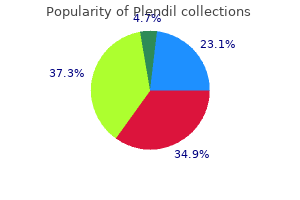
Buy generic plendil on-line. 100% blood pressure solution in marathi.
References
- Sheifer SE, Gersh BJ, Yanez ND III, et al: Prevalence, predisposing factors, and prognosis of clinically unrecognized myocardial infarction in the elderly. J Am Coll Cardiol 2000;35:119-120.
- Pendleton A, Arden N, Dougados M, et al. EULAR recommendations for the management of knee osteoarthritis: report of a task force of the Standing Committee for International Clinical Studies Including Therapeutic Trials (ESCISIT). Ann Rheum Dis 2000; 59(12):936-44.
- Tektonidou MG, Serelis J, Skopouli FN. Peripheral neuropathy in two patients with rheumatoid arthritis receiving infliximab treatment. Clin Rheumatol. 2007;26(2):258-260.
- Fraser DA, Bulat-Kardum L, Knezevic J, et al. Interferon-c receptor-1 gene polymorphism in tuberculosis patients from Croatia. Scand J Immunol 2003; 57: 480-484.
- Polascik TJ, Oesterling JE, Partin AW: Prostate specific antigen: a decade of discoveryowhat we have learned and where we are going, J Urol 162(2):293n306, 1999.
- Cousley RR, Calvert ML. Current concepts in the understanding and management of hemifacial microsomia. Br J Plast Surg 1997;50:536-551.
- Nelson LD: Continuous venous oximetry in surgical patients, Ann Surg 203:329, 1986.

